Home

atlet Înţelegere veteran mycoplasma microscope images extinde ști Aerodrom

Mycoplasma - UNC Lineberger

Detection of mycoplasma in contaminated mammalian cell culture using FTIR  microspectroscopy | SpringerLink
Detection of mycoplasma in contaminated mammalian cell culture using FTIR microspectroscopy | SpringerLink

Understanding mycoplasma testing

Figure 1 from Contamination of Tissue Cultures by Mycoplasmas | Semantic  Scholar
Figure 1 from Contamination of Tissue Cultures by Mycoplasmas | Semantic Scholar

Mycoplasma bacterial growth - Stock Video Clip - K005/7814 - Science Photo  Library
Mycoplasma bacterial growth - Stock Video Clip - K005/7814 - Science Photo Library

How to See Mycoplasmas

Detection and Antibiotic Treatment of Mycoplasma arginini Contamination in  a Mouse Epithelial Cell Line Restore Normal Cell Physiology
Detection and Antibiotic Treatment of Mycoplasma arginini Contamination in a Mouse Epithelial Cell Line Restore Normal Cell Physiology

PDF] Mycoplasma haemofelis infection and imaging of Mycoplasma haemofelis  using scanning electron microscopy in a cat. | Semantic Scholar
PDF] Mycoplasma haemofelis infection and imaging of Mycoplasma haemofelis using scanning electron microscopy in a cat. | Semantic Scholar
Frontiers | Differential Immunoreactivity to Bovine Convalescent Serum  Between Mycoplasma bovis Biofilms and Planktonic Cells Revealed by  Comparative Immunoproteomic Analysis
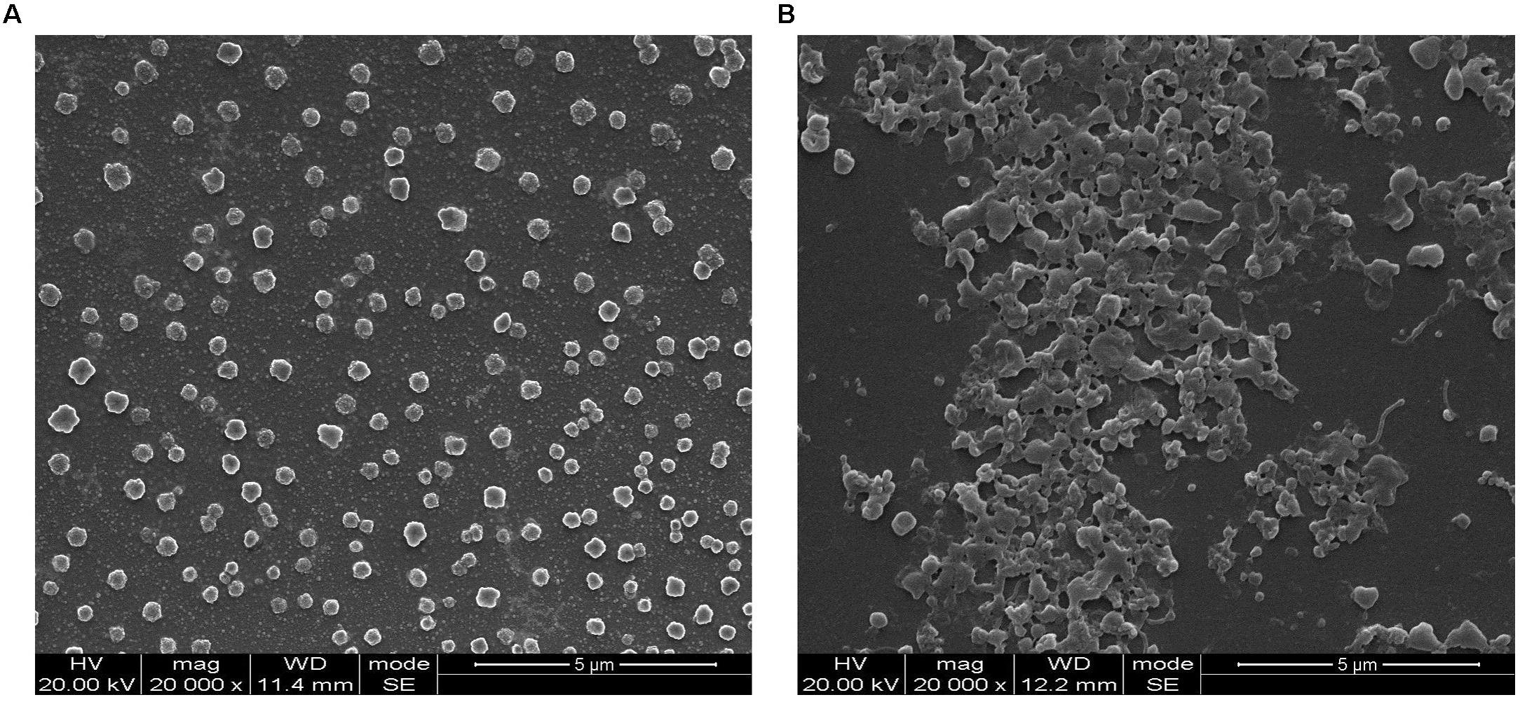
Frontiers | Differential Immunoreactivity to Bovine Convalescent Serum Between Mycoplasma bovis Biofilms and Planktonic Cells Revealed by Comparative Immunoproteomic Analysis

Mycoplasma - an overview | ScienceDirect Topics

Stereoscopic microscopy showing mycoplasma (A-B)and ureaplasma (C-D)... |  Download Scientific Diagram
Stereoscopic microscopy showing mycoplasma (A-B)and ureaplasma (C-D)... | Download Scientific Diagram

Microcolonies: a novel morphological form of pathogenic Mycoplasma spp |  bioRxiv
Microcolonies: a novel morphological form of pathogenic Mycoplasma spp | bioRxiv

Mycoplasma gallisepticum Inactivated by Targeting the Hydrophobic Domain of  the Membrane Preserves Surface Lipoproteins and Induces a Strong Immune  Response | PLOS ONE
Mycoplasma gallisepticum Inactivated by Targeting the Hydrophobic Domain of the Membrane Preserves Surface Lipoproteins and Induces a Strong Immune Response | PLOS ONE
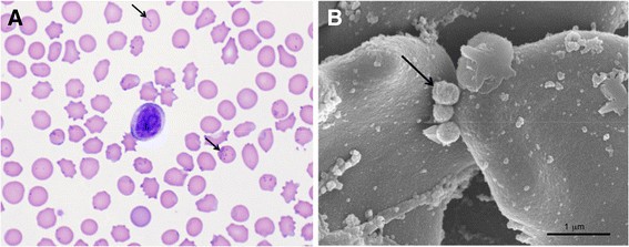
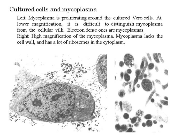
NIAH:NIAH Pathogenic Organisms Observed by Electron Microscope:Mycoplasma

Microscopy and genomic analysis of Mycoplasma parvum strain Indiana |  Veterinary Research | Full Text
Microscopy and genomic analysis of Mycoplasma parvum strain Indiana | Veterinary Research | Full Text

The atomic makeup of M. pneumoniae's 'nap' st | EurekAlert!

Using correlative microscopy for studying and treatment of Mycoplasma  infections of the ophtalmic mucosa
Using correlative microscopy for studying and treatment of Mycoplasma infections of the ophtalmic mucosa
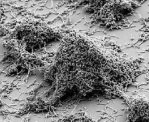
Cell Culture Basics - Mycoplasma 101 – A practical guide to prevention,  detection and elimination of mycoplasma contamination
Cell Culture Basics - Mycoplasma 101 – A practical guide to prevention, detection and elimination of mycoplasma contamination

Mycoplasmas and their host: emerging and re-emerging minimal pathogens:  Trends in Microbiology
Mycoplasmas and their host: emerging and re-emerging minimal pathogens: Trends in Microbiology

Mycoplasma pneumoniae: Disease-Pathogenesis for Labs | CDC

Scanning electron microscopy analysis on the Mycoplasma bovis 08M... |  Download Scientific Diagram
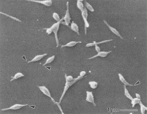
Scanning electron microscopy analysis on the Mycoplasma bovis 08M... | Download Scientific Diagram

American Society for Microbiology - Pic of the Day: Mycoplasma - adhesion  of symbiont infection structure to host This scanning electron micrograph  shows mycoplasma (colorized pink), a genus of bacteria that lack
American Society for Microbiology - Pic of the Day: Mycoplasma - adhesion of symbiont infection structure to host This scanning electron micrograph shows mycoplasma (colorized pink), a genus of bacteria that lack

SciELO - Brasil - Diagnosis and treatment of HEp-2 cells contaminated with  mycoplasma Diagnosis and treatment of HEp-2 cells contaminated with  mycoplasma
SciELO - Brasil - Diagnosis and treatment of HEp-2 cells contaminated with mycoplasma Diagnosis and treatment of HEp-2 cells contaminated with mycoplasma

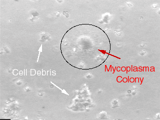
Microscopic image of Mycoplasma bovis colonies as fried eggs appearance...  | Download High-Quality Scientific Diagram
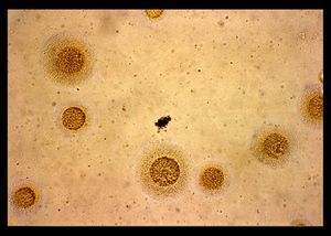
Microscopic image of Mycoplasma bovis colonies as fried eggs appearance... | Download High-Quality Scientific Diagram

Mycoplasma pneumoniae: Properties, Disease, Diagnosis – Microbe Online

Mycoplasma hominis - microbewiki

Morphologic features of Candidatus Mycoplasma haemohominis isolated... |  Download Scientific Diagram
Morphologic features of Candidatus Mycoplasma haemohominis isolated... | Download Scientific Diagram