Home

Deţinere culoare a sari the neuroglial cells microscope Simplu Păianjen web luptă

Microscopic Description -- Case 195

Neuron under Microscope with Labeled Diagram » AnatomyLearner >> The Place  to Learn Veterinary Anatomy Online
Neuron under Microscope with Labeled Diagram » AnatomyLearner >> The Place to Learn Veterinary Anatomy Online

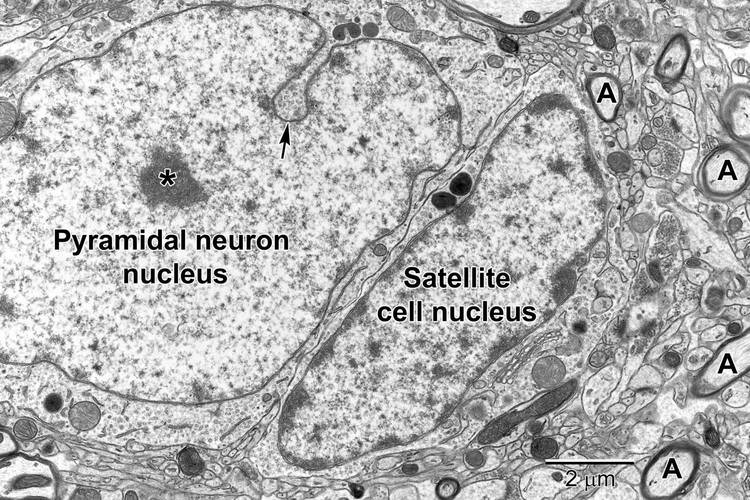
Frontiers | Brain Ultrastructure: Putting the Pieces Together

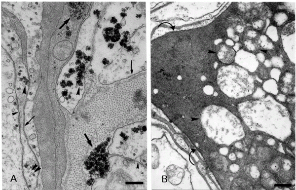
SciELO - Brasil - Electron microscopy of glial cells of the central nervous  system in the crab Ucides cordatus Electron microscopy of glial cells of the  central nervous system in the crab
SciELO - Brasil - Electron microscopy of glial cells of the central nervous system in the crab Ucides cordatus Electron microscopy of glial cells of the central nervous system in the crab
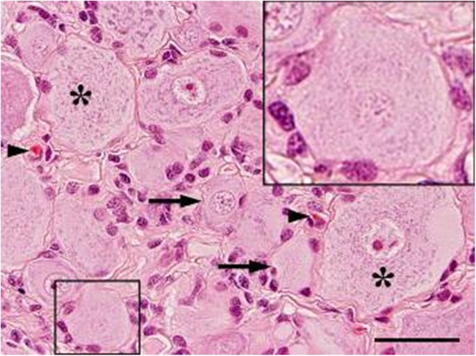
Canine dorsal root ganglia satellite glial cells represent an exceptional  cell population with astrocytic and oligodendrocytic properties |  Scientific Reports
Canine dorsal root ganglia satellite glial cells represent an exceptional cell population with astrocytic and oligodendrocytic properties | Scientific Reports

What are Glial Cells?

Element maps of glial cells. Light microscopy (LM) and element images... |  Download High-Quality Scientific Diagram
Element maps of glial cells. Light microscopy (LM) and element images... | Download High-Quality Scientific Diagram
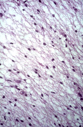
Brain glial cells, light micrograph - Oxford University Innovation

SciELO - Brasil - Electron microscopy of glial cells of the central nervous  system in the crab Ucides cordatus Electron microscopy of glial cells of the  central nervous system in the crab
SciELO - Brasil - Electron microscopy of glial cells of the central nervous system in the crab Ucides cordatus Electron microscopy of glial cells of the central nervous system in the crab

Neuron under Microscope with Labeled Diagram » AnatomyLearner >> The Place  to Learn Veterinary Anatomy Online
Neuron under Microscope with Labeled Diagram » AnatomyLearner >> The Place to Learn Veterinary Anatomy Online

Glial cells, SEM - Stock Image - C003/5964 - Science Photo Library

Glial cells: Histology and clinical notes | Kenhub

Nervous System Lab
Nervous System Lab

Astrocytoma at 40x Magnification | Nikon's MicroscopyU

Central Nervous System | histology

Mammal. Spinal cord. Neuron. 500X - Neuron - Mammals - Mammals - Nervous  system - Other systems - Comparative anatomy of Vertebrates - Animal  histology - Photos
Mammal. Spinal cord. Neuron. 500X - Neuron - Mammals - Mammals - Nervous system - Other systems - Comparative anatomy of Vertebrates - Animal histology - Photos

Microscopic lesions and positive viral antigen cells in naturally... |  Download Scientific Diagram
Microscopic lesions and positive viral antigen cells in naturally... | Download Scientific Diagram

Light Micrograph of Human Brain Tissue Showing Neurons and Glial Cells  Stock Image - Image of stain, healthy: 109142315
Light Micrograph of Human Brain Tissue Showing Neurons and Glial Cells Stock Image - Image of stain, healthy: 109142315

Neurons and glial cell types in area 32 of the monkey cerebral... |  Download Scientific Diagram
Neurons and glial cell types in area 32 of the monkey cerebral... | Download Scientific Diagram

Satellite glial cell - Wikipedia

Lab 1 Neurohistology - Neuroglia